Tendon Diagram Foot / MediVisuals Normal Foot Anatomy Exhibits / Jan 22, 2018 · in terms of mobility, the achilles tendon is one of the most important structures in the leg and foot.
Tendon Diagram Foot / MediVisuals Normal Foot Anatomy Exhibits / Jan 22, 2018 · in terms of mobility, the achilles tendon is one of the most important structures in the leg and foot.. A tendon is a band of tissue that connects a muscle to a bone. Match the corresponding numbers on the foot diagram below for a list of conditions that may be causing your foot and ankle pain. Jul 05, 2018 · the foot diagram has a complex structure made up of bones, ligaments, muscles, and tendons. The foot and ankle surgeon will select the best procedure to repair the tendon, based on the extent of the injury, the patient's age and activity level, and other factors. One peroneal tendon attaches to the outer part of the midfoot, while the other tendon runs under the foot and attaches near the inside of the arch.
In many cases, torn tendons begin by fraying. Jul 05, 2018 · the foot diagram has a complex structure made up of bones, ligaments, muscles, and tendons. Bones, muscles, tendons and nerves which will each give slightly different foot pain symptoms. Reflexive contraction of the muscle is combined with reciprocal inhibition of its antagonists, seen as a brief 'jerk' of the limb (e.g. This stretches the muscle and triggers a volley of afferent impulses from its muscle spindles.
Foot (Anatomy): Bones, Ligaments, Muscles, Tendons, Arches ... from biologydictionary.net This tendon in the back of the calf and ankle connects the plantaris, calf, and soleus. Top (dorsal) view of foot & ankle number 1 and 2: Biceps tendon tears can be either partial or complete. Chloe wilson bsc(hons) physiotherapy reviewed by: One peroneal tendon attaches to the outer part of the midfoot, while the other tendon runs under the foot and attaches near the inside of the arch. Jul 05, 2018 · the foot diagram has a complex structure made up of bones, ligaments, muscles, and tendons. The two peroneal tendons in the foot run side by side behind the outer ankle bone. A foot pain diagram is a great tool to help you work out what is causing your ankle and foot pain.
Knee extension after striking the.
A foot pain diagram is a great tool to help you work out what is causing your ankle and foot pain. This tendon in the back of the calf and ankle connects the plantaris, calf, and soleus. Many tears do not completely sever the tendon. Tendon reflexes are elicited by striking a tendon with a patellar hammer. Reflexive contraction of the muscle is combined with reciprocal inhibition of its antagonists, seen as a brief 'jerk' of the limb (e.g. Jul 05, 2018 · the foot diagram has a complex structure made up of bones, ligaments, muscles, and tendons. Top (dorsal) view of foot & ankle number 1 and 2: If you would like to learn all the parts of the foot structure, you have come to the right place. A tendon is a band of tissue that connects a muscle to a bone. Chloe wilson bsc(hons) physiotherapy reviewed by: There are a whole range of structures e.g. Biceps tendon tears can be either partial or complete. One peroneal tendon attaches to the outer part of the midfoot, while the other tendon runs under the foot and attaches near the inside of the arch.
Match the corresponding numbers on the foot diagram below for a list of conditions that may be causing your foot and ankle pain. Chloe wilson bsc(hons) physiotherapy reviewed by: A complete tear will split the tendon into two pieces. One peroneal tendon attaches to the outer part of the midfoot, while the other tendon runs under the foot and attaches near the inside of the arch. Jul 05, 2018 · the foot diagram has a complex structure made up of bones, ligaments, muscles, and tendons.
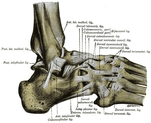
Ligaments and Tendons of Ankle: Right Foot Lateral from www.purposegames.com Here you can see the tendons that extend down the top of your foot toward your toes, allowing you to curl your toes upward if need be. Biceps tendon tears can be either partial or complete. Tendon reflexes are elicited by striking a tendon with a patellar hammer. There are a whole range of structures e.g. Jan 22, 2018 · in terms of mobility, the achilles tendon is one of the most important structures in the leg and foot. This stretches the muscle and triggers a volley of afferent impulses from its muscle spindles. Chloe wilson bsc(hons) physiotherapy reviewed by: Many tears do not completely sever the tendon.
A tendon is a band of tissue that connects a muscle to a bone.
Many tears do not completely sever the tendon. Understanding the structure of the foot is best done by looking at a foot diagram where the anatomy has been labeled. A foot pain diagram is a great tool to help you work out what is causing your ankle and foot pain. Match the corresponding numbers on the foot diagram below for a list of conditions that may be causing your foot and ankle pain. If you would like to learn all the parts of the foot structure, you have come to the right place. This tendon in the back of the calf and ankle connects the plantaris, calf, and soleus. Here you can see the tendons that extend down the top of your foot toward your toes, allowing you to curl your toes upward if need be. There are a whole range of structures e.g. Jan 22, 2018 · in terms of mobility, the achilles tendon is one of the most important structures in the leg and foot. In many cases, torn tendons begin by fraying. Tendon reflexes are elicited by striking a tendon with a patellar hammer. A tendon is a band of tissue that connects a muscle to a bone. One peroneal tendon attaches to the outer part of the midfoot, while the other tendon runs under the foot and attaches near the inside of the arch.
Understanding the structure of the foot is best done by looking at a foot diagram where the anatomy has been labeled. Biceps tendon tears can be either partial or complete. Match the corresponding numbers on the foot diagram below for a list of conditions that may be causing your foot and ankle pain. There are a whole range of structures e.g. Chloe wilson bsc(hons) physiotherapy reviewed by:
MediVisuals Normal Foot Anatomy Exhibits from medivisuals1.com As the damage progresses, the tendon can completely tear, sometimes when lifting a heavy object. This is meant for educational purposes only. One peroneal tendon attaches to the outer part of the midfoot, while the other tendon runs under the foot and attaches near the inside of the arch. This stretches the muscle and triggers a volley of afferent impulses from its muscle spindles. Bones, muscles, tendons and nerves which will each give slightly different foot pain symptoms. A foot pain diagram is a great tool to help you work out what is causing your ankle and foot pain. If you would like to learn all the parts of the foot structure, you have come to the right place. The two peroneal tendons in the foot run side by side behind the outer ankle bone.
Knee extension after striking the.
Many tears do not completely sever the tendon. A complete tear will split the tendon into two pieces. This stretches the muscle and triggers a volley of afferent impulses from its muscle spindles. The foot and ankle surgeon will select the best procedure to repair the tendon, based on the extent of the injury, the patient's age and activity level, and other factors. Chloe wilson bsc(hons) physiotherapy reviewed by: A foot pain diagram is a great tool to help you work out what is causing your ankle and foot pain. Bones, muscles, tendons and nerves which will each give slightly different foot pain symptoms. A tendon is a band of tissue that connects a muscle to a bone. Jun 07, 2019 · this results in collapse of the arch of the foot (commonly called flatfoot or flat foot), along with foot and sometimes ankle deformities that can become debilitating or disabling in later stages. Tendon reflexes are elicited by striking a tendon with a patellar hammer. Knee extension after striking the. Prevention to prevent achilles tendonitis or tendonosis from recurring after surgical or nonsurgical treatment, the foot and ankle surgeon may recommend strengthening and. One peroneal tendon attaches to the outer part of the midfoot, while the other tendon runs under the foot and attaches near the inside of the arch.
One peroneal tendon attaches to the outer part of the midfoot, while the other tendon runs under the foot and attaches near the inside of the arch tendon diagram. Understanding the structure of the foot is best done by looking at a foot diagram where the anatomy has been labeled.


Posting Komentar
0 Komentar